
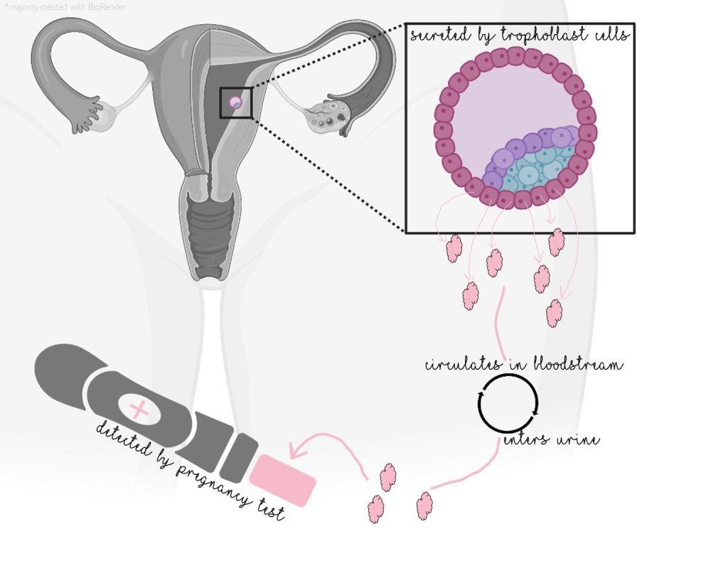
Pregnancy can be detected by looking for the glycoprotein human chorionic gonadotropin, which functions as a hormone. After an egg has been successfully fertilized, the resulting cell multiplies into a mostly hollow ball of cells called a blastula. Trophoblast cells, the cells on the exterior of the blastula, make and secrete hCG when the blastula implants in the uterus. Then hCG levels continue to increase for the first eight weeks, staying up for the rest of the pregnancy.
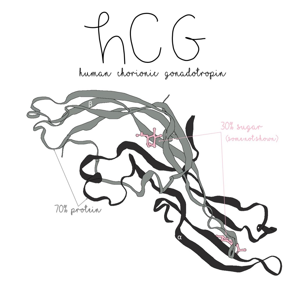
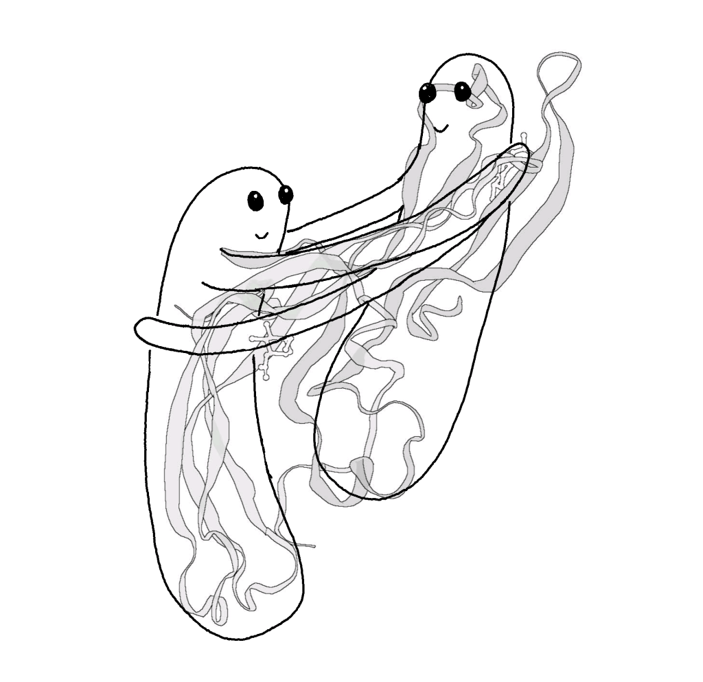
hCG is a glycoprotein. Two protein subunits, named alpha and beta, make up about 70% of its mass while sugars make up the remaining 30%. This is what it looks like:
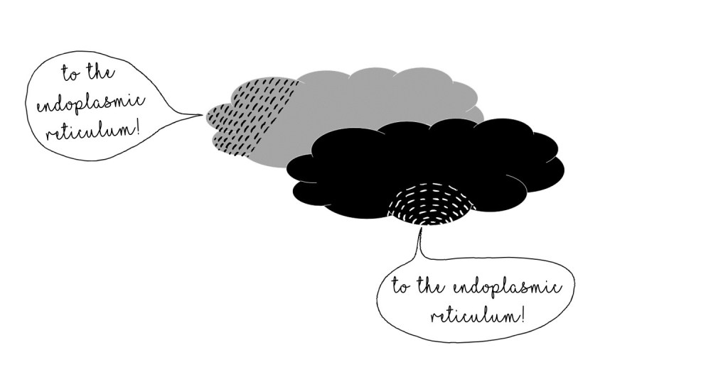
The protein parts of hCG are built at ribosomes in the cell’s rough endoplasmic reticulum (ER). Each subunit starts out as a longer protein with extra pieces, pre-alpha and pre-beta. These extra pieces, called signal peptides, tell other parts of the cell where the protein needs to go.
Building the subunits is only one part of making a protein. A lot of other stuff is going on while the protein is being put together:
- The signal peptides are removed (after each part gets to its destination, of course)
- Some sugars are added on
- Disulfide bridges form to stabilize the protein’s structure

The two subunits join somewhat like a hug–one disulfide bridge even locks them together!
After all of this, hCG is transferred to the Golgi to get even more sugars tacked on before it’s ready for secretion.
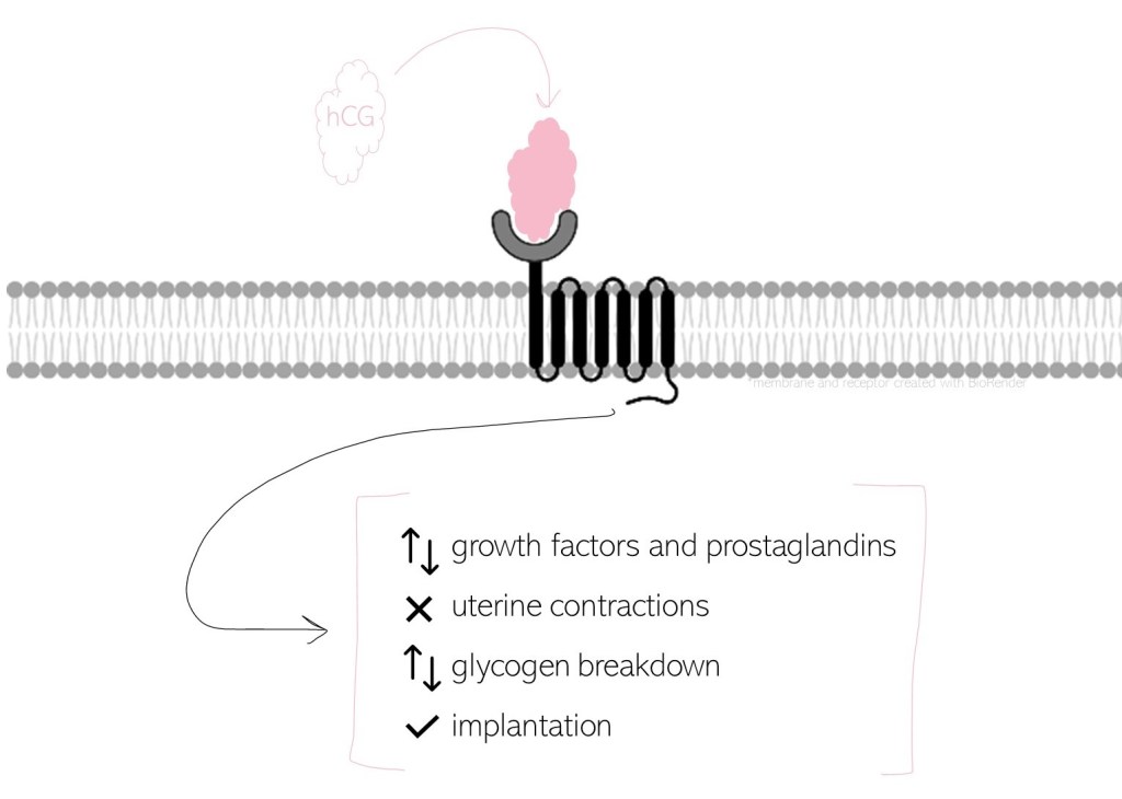
Since hCG is a hormone, it circulates throughout the body triggering different parts to do something. hCG…
- regulates the production of other hormones like growth factors and prostaglandins
- inhibits uterine contractions
- influences the breakdown of glycogen (stored sugar)
- somewhat coordinates implantation
Certainly hCG is a critical hormone that synchronizes all kinds of necessary changes at the start of a pregnancy–including letting you know about it!
Wu, et al. Structure of human chorionic gonadotropin at 2.6 A resolution from MAD analysis of the selenomethionyl protein, Structure, Volume 2, Issue 6, Pages 545-558, (Free PDF: https://www.cell.com/structure/pdf/S0969-2126(00)00054-X.pdf) (on PDB as 1HCN)
S.F. de Medeiros, R.J. Norman, Human choriogonadotrophin protein core and sugar branches heterogeneity: basic and clinical insights, Human Reproduction Update, Volume 15, Issue 1, January-February 2009, Pages 69–95, https://doi.org/10.1093/humupd/dmn036
https://www.yourhormones.info/hormones/human-chorionic-gonadotrophin/

One Reply to “”