
Green Fluorescent Protein goes by the nickname “GFP.” It was first found in jellyfish and named for its unique ability to fluoresce, or emit light, that is–you guessed it–the color green!

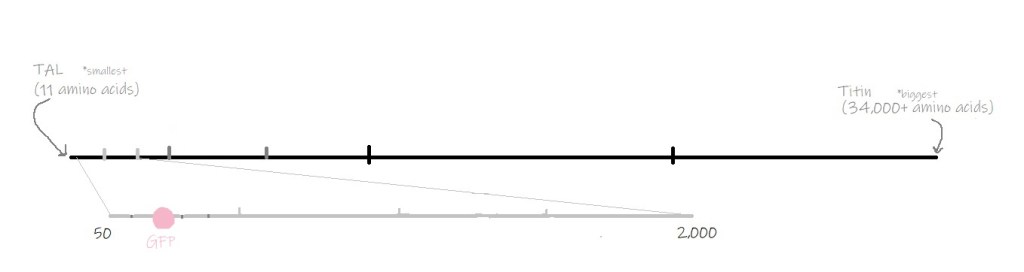
GFP is made of 236 amino acids (protein building blocks) that form what is known as a beta-barrel structure.
Inside of the beta barrel lies a few very special amino acids that come together to form a fluorophore. A fluorophore is a molecule that emits light when it is activated by other light. For example, when GFP’s fluorophore is activated by UV or blue light, it emits green light. Because of this, GFP’s structure has been described fittingly as “a candle in a lantern.”

After Dr. Osamu Shimomura discovered GFP, Dr. Martin Chalfie revolutionized biological research by using GFP to label other proteins in live cells. Proteins are far too small to be seen under a typical microscope, so to study proteins in cells, they must be labeled with some sort of marker. Before GFP, the only ways scientists could label proteins inside of cells would kill the cells.
Instructions in the DNA control the production of all proteins in a cell, including GFP. Dr. Chalfie figured out that he could combine the instructions for GFP with the instructions for a protein of interest to tell cells to make a fusion protein. Effectively, this “glued” GFP onto another protein within the cell. With GFP attached, Dr. Chalfie could track a protein of interest inside of a living cell.
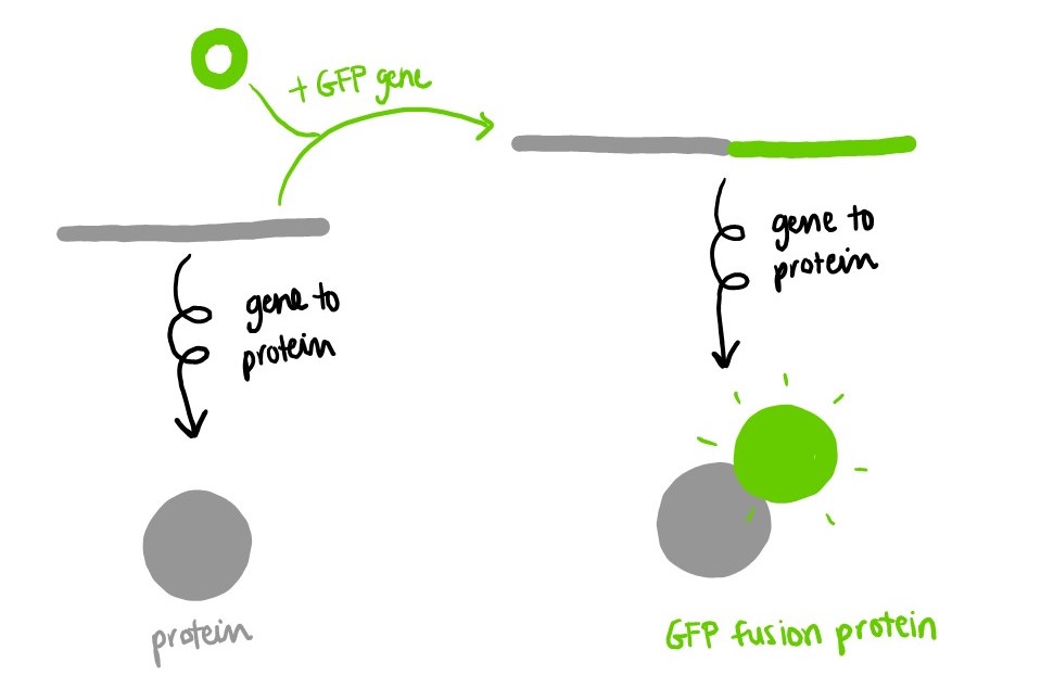
Today, thousands of scientists around the world use GFP to investigate proteins inside of living cells and even whole organisms! We can watch these proteins move around, seeing where they go and where they came from. GFP helped change our picture of proteins in the cell from a static photo to an action-packed movie.
This discovery was so important and revolutionary that the two scientists mentioned above were awarded a Nobel Prize along with Dr. Roger Tsien, who studied how GFP worked and engineered other versions that emit different colors of light.

For more about how GFP transformed scientific research: How glow-in-the-dark jellyfish inspired a scientific revolution
*Dr. Chalfie didn’t come up with the idea of tagging proteins with GFP first. See this article for more about who did: https://www.sciencemag.org/careers/2009/02/man-who-wasnt-there
Barondeau, D. P., Putnam, C. D., Kassmann, C. J., Tainer, J. A., & Getzoff, E. D. (2003). Mechanism and energetics of green fluorescent protein chromophore synthesis revealed by trapped intermediate structures. Proceedings of the National Academy of Sciences of the United States of America, 100(21), 12111–12116. https://doi.org/10.1073/pnas.2133463100

2 Replies to “Meet Green Fluorescent Protein!”