
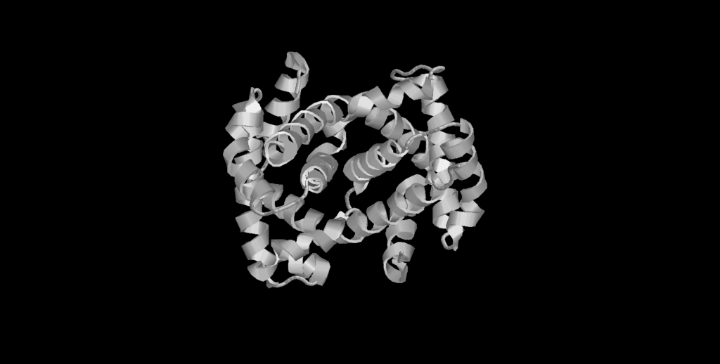
If you look at any of my posts introducing a specific protein, you will always see an image like this one:
Maybe you see these images and ask,
“What are all these squiggly lines you keep showing us?”
These squiggly lines are three-dimensional representations of proteins. Every protein has a structure, and its structure is critical to its function. It is important for scientists and bioengineers to know what a protein “looks like” so that we can learn more about how it does what it does.
People used to show protein structures with drawings and models like these:
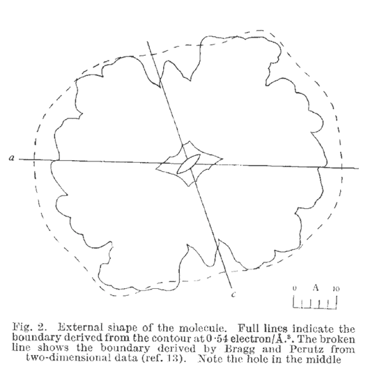
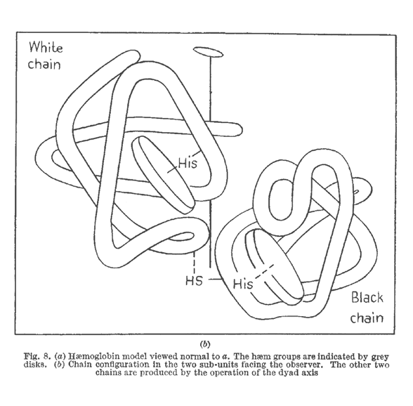
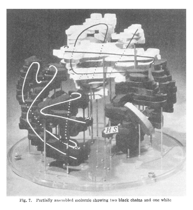
These representations can be somewhat useful if studied carefully, but it can be difficult to find patterns this way.
Luckily, biophysicist Jane Richardson found a much better way to draw these protein structures!

In the 1980s, Jane Richardson began sketching proteins in a new, elegantly simple way now known as a “Richardson diagram” or “ribbon diagram.” Because these are more simple, they allow scientists to find patterns and motifs much more easily.
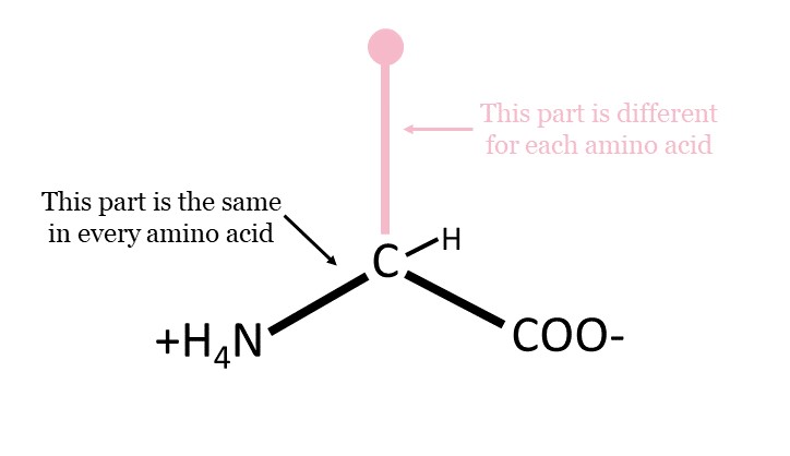
The basis of a ribbon diagram’s style depends on secondary structure. Remember, a protein is made from a chain of pieces called amino acids. Each amino acid has a portion that is unique and a portion that is the same.
The secondary structure of a protein depends on interactions between those portions of amino acids that are all the same, and it usually looks like one of two things: an alpha helix or a beta sheet.
Alpha helices
An alpha helix looks like a spring. The unique portions of its amino acids stick out away from the center, while the same portions interact with each other along the axis of the spring.

Beta sheets
In a beta sheet, the unique portions of the amino acids stick out either up or down, while the same portions interact with each other side-to-side. Arrows indicate the direction of the beta sheet and point towards the end of the chain.

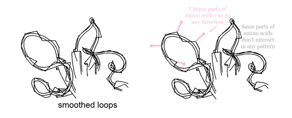
Linkers
Not everything can have secondary structure though! Proteins also need flexible pieces to link things together. In a ribbon diagram, linkers are smoothed into curves. Note that “linkers” don’t necessarily have to link anything together. It’s just that the orientations of their amino acids don’t have any particular pattern.
Since hand-drawings are difficult to share and manipulate (and not all scientists can draw well!), computer programs have been developed to model ribbon diagrams based off of Jane Richardson’s design. When scientists find the structure of a protein, they upload a .pdb file onto the Protein Data Bank to share it with others, and each new structure gets a 4-character accession number.
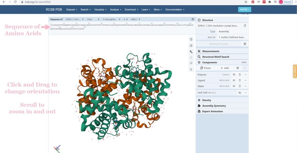
I use the PDB on a regular basis in my work, but the PDB is not just for scientists. You can look up all of these structures in the PDB too! And it’s actually incredibly easy. I’ll show you how to do it for our example, hemoglobin.
- Visit https://www.rcsb.org/ (This is where the PDB lives) and use the search bar to… well, search!
- Select the structure you want. Each entry will give information about how the structure was determined, the resolution of the structure (the smaller number, the better), an R-value (somewhat describes how flexible the protein is), and more.
- Clicking on the “3D View” tab will bring you to a 3D viewer where you can move the protein model around, zoom in (using the scroll wheel on your mouse), and look at it in detail. Scientists often have other 3D viewing programs with more bells and whistles, but all of this can be done right in the browser, no extensions or external software needed!


Thank you to Jane Richardson for using your brains and your art to change protein structure representation for the better!
So these structures can look cool, even beautiful at the right angles, but how do we find these structures? How do we know what a protein looks like? That’s a big question for another time (future post hopefully?), but for now, check out these resources to learn more about how we figure it out:
- X-ray crystallography
- Cryo-EM
PERUTZ, M., ROSSMANN, M., CULLIS, A. et al. (1960). Structure of Hæmoglobin: A Three-Dimensional Fourier Synthesis at 5.5-Å. Resolution, Obtained by X-Ray Analysis. Nature, 185, 416–422. https://doi.org/10.1038/185416a0
For Jane Richardson’s drawings: https://commons.wikimedia.org/wiki/User:Dcrjsr/gallery_of_protein_structure

One Reply to “”